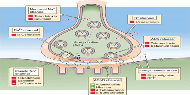
Structure activity relationship of Synaptic & Junctional Transmission

Synaptic transmission: functional anatomy types of synapses
The anatomic structure of synapses varies considerably in the different parts of the mammalian nervous system. The ends of the presynaptic fibers are generally enlarged to form terminal buttons. In the cerebral and cerebellar cortex, endings are commonly located on dendrites and frequently on dendritic spines, small knobs projecting from dendrites. In some instances, the terminal branches of the axon of the presynaptic neuron form a basket or net around the soma of the postsynaptic cell (basket cells of the cerebellum and autonomic ganglia). In other locations, they intertwine with the dendrites of the postsynaptic cell (climbing fibers of the cerebellum) or end on the dendrites directly (apical dendrites of cortical pyramidal cells).
Presynaptic & postsynaptic structure & function
Each presynaptic terminal of a chemical synapse is separated from the postsynaptic structure by a synaptic cleft that is 20 to 40 nm wide. Across the synaptic cleft are many neurotransmitter receptors in the postsynaptic membrane, and usually, a postsynaptic thickening called the postsynaptic density. The postsynaptic density is an ordered complex of specific receptors, binding proteins, and enzymes induced by postsynaptic effects. Inside the presynaptic terminal are many mitochondria and many membrane-enclosed vesicles, which contain neurotransmitters.
Excitatory & inhibitory postsynaptic potentials
The puncture of a cell membrane is signaled by the appearance of a steady 70-mV potential difference between the microelectrode and an electrode outside the cell. The cell can be identified as a spinal motor neuron by stimulating the appropriate ventral root and observing the cell’s electrical activity.
Temporal & spatial summation
Summation may be temporal or spatial. Temporal summation occurs if repeated afferent stimuli because new EPSPs before previous EPSPs have decayed. A longer time constant for the EPSP allows for a more incredible opportunity for the outline. When activity is present in more than one synaptic knob simultaneously, spatial summation occurs, and training in one synaptic knob summates with activity in another to approach the firing level. THEREFORE, the EPSP is not an all-or-none response but is proportionate in size to the strength of the afferent stimulus.
Slow postsynaptic potentials
In addition to the EPSPs and IPSPs described previously, slow EPSPs and IPSPs have been expressed in autonomic ganglia, cardiac and smooth muscle, and cortical neurons. These postsynaptic potentials have a latency of 100 to 500 ms and last several seconds. The slow EPSPs are generally due to decreases in K+ conductance, and the slow IPSPs are due to increases in K+ conductance.
Generation of the action potential in the postsynaptic neuron
The constant interplay of excitatory and inhibitory activity on the postsynaptic neuron produces a fluctuating membrane potential, the algebraic sum of the hyperpolarizing and depolarizing activity. The soma of the neuron thus acts as a sort of integrator. When the 10 to 15 mV of depolarization sufficient to reach the firing level is attained, a propagated spike results. However, the discharge of the neuron is slightly more complicated than this. In motor neurons, the portion of the cell with the lowest threshold for producing a fullfledged action potential is the initial segment, the amount of the axon at and just beyond the axon hillock.
Electrical transmission
At synaptic junctions where transmission is electrical, the impulse reaching the presynaptic terminal generates an EPSP in the postsynaptic cell that has a much shorter latency than the low-resistance bridge between the two EPSP at a synapse where transmission is chemical. A chemically mediated postsynaptic response occurs in conjoint synapses in both a short-latency reaction and a longer-latency.
Inhibition & facilitation at synapses
Inhibition in the CNS can be postsynaptic or presynaptic. Postsynaptic inhibition during an IPSP is called direct inhibition because it is not a consequence of previous discharges of the postsynaptic neuron. There are various forms of indirect inhibition, which is inhibition due to the effects of the last postsynaptic neuron discharge. For example, the postsynaptic cell can be refractory to excitation because it has just fired and is in its refractory period. After hyperpolarization, it is also less excitable. This after-hyperpolarization may be significant and prolonged in spinal neurons, especially after repeated firing.
Summary
Various pathways in the nervous system mediate postsynaptic inhibition, and one illustrative example is presented here. Afferent fibers from the muscle spindles (stretch receptors) in skeletal muscle project directly to the spinal motor neurons of the motor units supplying the same muscle. Impulses in this afferent fiber cause EPSPs and, with the summation, propagated responses in the postsynaptic motor neurons. At the same time, IPSPs are produced in motor neurons supplying the antagonistic muscles, which have an inhibitory interneuron interposed between the afferent fiber and the motor neuron.


